Pisum Genetics
2006—Volume 38
Research Papers
A new dominant-acting necrosis mutation in pea
S. M. Rozov
Inst. of Cytol. and Genet. Siberian Branch of Russ. Acad. of Sci., Novosibirsk, Russia
The necrosis phenotype in pea consists of a relatively small class of mutations. The majority of these mutations produce small necrotic spots or marginal stripes on leaves and stipules. The spots and stripes may be of different color — from yellow to orange or dark brown. In some mutants the spots can enlarge and form necrotic sectors on the leaves. The well-known necrosis mutations are: nec (6), necrosis of leaf and stipule margins, that covers the intervenial area as well as the veins; len (4), leaf-edge necrosis, that forms a necrotic spots or areas at the leaflet margins; gn (7), gold necrosis, which forms the gold orange spots and areas on underside of the leaflets; brz or dgl (2,3), bronze or degenerating leaves, which forms deep-yellow to dark-brown growing necrotic spots or areas on older leaves and stipules, caused by the excessive iron accumulation in some cells of pea shoot tissues (1); and bulf (5), burnt leaf, that causes brown necrotic stripes on the periphery of leaflets and stipules. All of these mutations are recessive in nature. In the present work I describe a dominant EMS-induced mutation associated with leaf necrosis. During the screening of an M2 progeny of the EMS-treated line SGE, the SGE–1002 mutant was isolated. This mutation is characterized by the presence of dry, pale-yellow sectors on leaflets and stipules. Tissues in these areas look like leaf lamina of the mature pea plants that have naturally senesced (Fig. 1, 2).
The dry necrotic lesions on leaflets and stipules in the SGE–1002 mutant line appear very quickly and spontaneously: one day the leaflets or stipules are normal and green, without any visible defects, the next day the almost dry pale-yellowish sectors are present. Dry necrotic sectors in the SGE–1002 mutants can
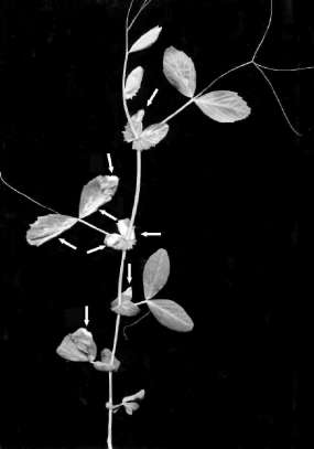
Fig. 1. A plant of SGE–1002 line, showing the dry necrosis phenotype. Arrows point to the dry pale-yellowish sectors of the naturally senesced tissues.
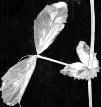
Fig.2. A leaf of the SGE–1002 mutant. The dry necrotic sectors are clearly visible as the light areas.
29