PNL Volume 21 1989 RESEARCH REPORTS                       77
COMPARATIVE STUDIES OF THE SPONTANEOUS AND INDUCED VARIABILITY IN CALLUS CULTURES AND RECENERATED PLANTS OF PEA
Tikhvinskaya, N. S. and S. A. Gostimski
Genetics Department, Moscow State University, Moscow 117234, USSR
It is supposed that different mutagens may significantly influence the frequency and spectrum of mutations in plant tissue culture systems. Evaluation of induced variability must be considered in relation to soma-clonal variability in tissue culture (3).
The comparison was made between spontaneous and X-ray induced varia­bility in long-term callus cultures and regenerated plants (R0) of pea lines 17-35 and 18-1. The callus lines used in the experiments originated from apices of plants heterozygous for yellow xantha mutations. In each line the mutant phenotype was regulated by one nuclear gene. In line 18-1 the mutation was semidominant and in the heterozygous state caused a light-green colour of the pea leaves. The use of a marker line allowed us to observe the somatic mutations which manifested as yellow or twin yellow/dark-green spots or sectors on the leaves of the plants. The muta tion in line 17-35 was recessive and somatic mutations could be observed as yellow sectors.
Callus tissue was cultured on B5 medium (2) supplemented with NAA (0.1 mg/l) and BAP (0.5 mg/l). The pieces of callus (4-5 mm2) were X-irradiated with doses of 1, 2.5, 5, 10, and 20 kr (dose rate 1.5 kr/min). After treatment the callus tissues were transferred to the regenerating medium with 5 mg/l BAP. Regenerated shoots were placed in tubes on 1/2 B5 medium where the plants were grown to the flowering stage. We ob­served alterations in plant morphology and counted somatic mutant sectors on the leaves of regenerated plants.
For measurement of the radiosensitivity of callus the "mitotic index" of the tissue 48 h after treatment and the "growth factor" were used. The growth factor is defined as the ratio of final fresh weight of callus : initial weight. It was shown that doses of 2.5 and 5 kr greatly suppressed cell division in pea callus. At high X-ray doses of 10 and 20 kr cell division was completely inhibited and all pieces of callus died within the two weeks following irradiation. X-rays at 1 kr did not signi­ficantly influence callus growth of either line and the growth factors were close to those for untreated controls. However, at 2.5 and 5 kr growth was essentially decreased (Fig. 1). Bud formation in non-irra­diated controls and the 1 kr treatment was observed at the end of the-third week on regenerating medium. Doses of 2.5 and 5 kr significantly inhibited bud formation in both callus lines 17-35 and 18-1. The average number of shoots per callus was reduced with the increase in radiation dose (Fig. 2).
The number of altered regenerated plants increased with the rise in X-ray dose. The character of the alteratons was the same for the control and irradiated material and related to plant habit and stem and leaf mor­phology. In addition, we observed the appearance of xantha mutants (Table 1). The frequency of somatic mutant sectors on the leaves of R0 re­generated plants also increased for both lines with higher X-ray doses (Table 2). In the non-irradiated control the frequency of mutant sectors
78
PNL Volume 21 1989 RESEARCH REPORTS
increased fivefold in comparison with that of plants grown from seeds in field experiments. For all X-ray doses the incidence of somatic sectors did not exceed the values obtained in field. The events of mitotic crossingover ,(the origin of twin spots) were registered only in the un­treated control of line 18-1.
Thus we evaluated the radiosensitivity of callus cultures of two pea lines 17-35 and 18-1. The results show that morphogenic pea callus lines heterozygous for yellow xantha mutations were very suitable for the inves­tigation of mutagenic effects induced by different X-ray doses. The sys­tem provided an opportunity to register homozygous chlorophyll mutations. Alterations among the R0 regenerated plants arose both in the control and at all doses of X-rays. Irradiation did not change the range of phenotypic variants. We may conclude that somaclonal variation is possi­bly not qualitatively different from that induced by X-rays. The same conclusion was made by Novak et al . (1) concerning chemical mutagenesis in vitro for maize.
1.   Evola, S. V., F. A. Burr, and B. Burr. 1984. Plant Mol. Biol, (abstract of the Eleventh Aharon Katzir-Katchalsky Conference), Jerusalem.
2.   Gamborg, 0. L., R. A. Miller, and K. Ojima. 1968. Exp. Cell Res. 50:151-158.
3.   Novak, F. J., R. Afza, S. Daskalov, T. Hermelin, and T. Lucretti. 1986. In: Nuclear Techniques and in vitro Culture for Plant Improvement. Vienna. pp. 29-33.
4.   Novak, F. J., T. Hermelin, S. Daskalov, and M. Nesticky. 1986. In: Genetic Manipulation in Plant Breeding. Proc. Int. Symp. (Berlin) 1985. pp. 564-576.
Table 1. Percentage of X-ray-induced 'xantha' mutants in regenerated plants of pea lines 18-1 and 17-35.
Dose (kr)
Number of regenerated plants
Frequency of chlorophyll mutants (%)
18-1
Control
314
0.32
1.0
384
1.04
2.5
127
1.57
5.0
21
0.00
17-35
Control
310
0.32
1.0
66
0.00
2.5
49
4.10
5.0
32
0.00
PNL Volume 21 1989 RESEARCH REPORTS
79
Table 2. Influence of X-ray doses on the incidence of altered RQ
regenerated plants and on the frequency of somatic mutant sectors in lines 18-1 and 17-35.
Dose (kr)
Number of regenerated plants
Altered plants (%)
Frequency of mutant sectors, %
yellow aa
dark-green AA
twin AA/aa
18-1
Control
103
27.0
4.05
1 .35
2.70
1.0
119
28.3
7.08
0.88
0.00
2.5
96
46.3
7.25
0.00
0.00
5.0
21
61.5
7.69
0.00
0.00
17-35
Control
118
29.7
2.54
0.00
0.00
1 .0
34
26.5
0.00
0.00
0.00
2.5
42
50.0
2.40
0.00
0.00
5.0
32
37.5
6.25
0.00
0.00
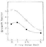
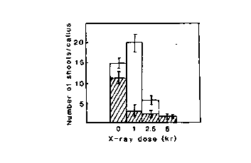
Fig. 1. Influence of the radiation doze on the callus growth rate of lines 17-35 (square) and 18-1 (circle) in vitro
Fig. 2. Avarage number of regenerated shoots per X-irradiated callus of lines 17-35 (empty bars) and 18-1 (filled bars).