During pea seed development a mass of storage proteins is synthe-
sized and accumulated within the cotyledon parenchyma cells (1). This
biosynthesis is correlated with dramatic changes in the subcellular or-
ganization of the storage parenchyma cells.
In order to get a general view of the morphological characteristics
at different developmental stages, a series of light microscopic studies
was performed. Plants of Pisum sativum cv. 'Dippes Gelbe Viktoria' were
grown in a greenhouse under controlled conditions and pods were harvested
13, 15, 17, 20, 23, 27, and 31 days after flowering. The ripening seeds
were fixed with glutaraldehyde, embedded in Spurr's resin, and semin-thin
sections were cut.
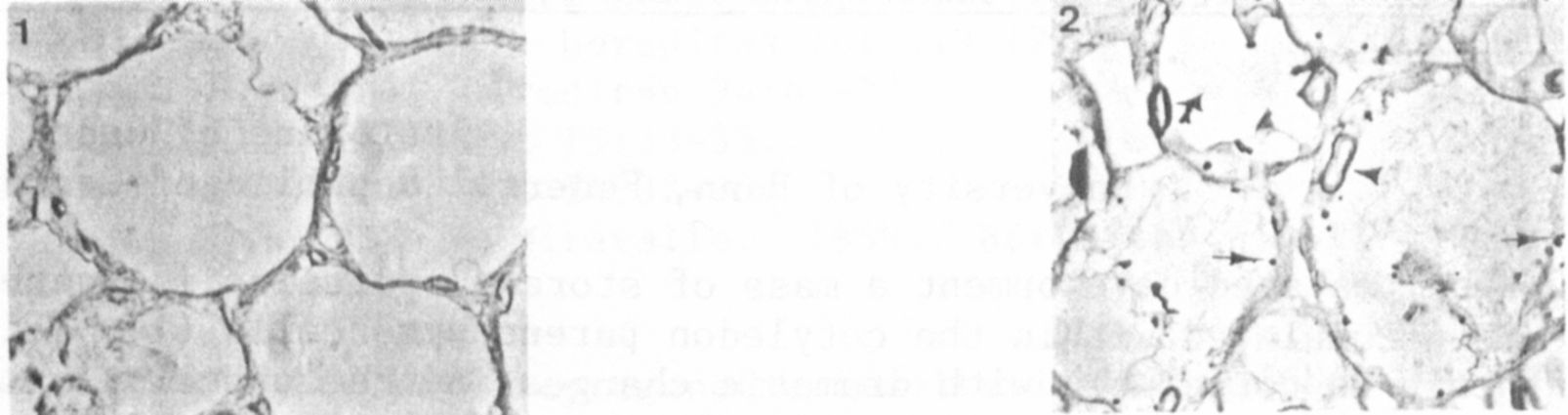
Cell divisions in the cotyledon parenchyma tissue had ceased 13 days
after flowering. At this time the parenchyma cells contained a single
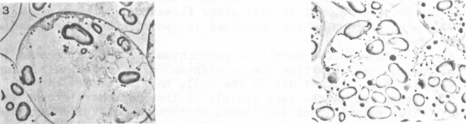
large vacuole (Fig. 1). At day 15 the cells had increased in size
(Fig. 2). Dense aggregates were visible at the periphery of the vacuoles
(arrows). This material has been immunohistochemically identified as
storage protein by Craig and Goodchild (2). A few starch grains were
detectable in the cytoplasm (arrowheads). The vacuoles of some cells
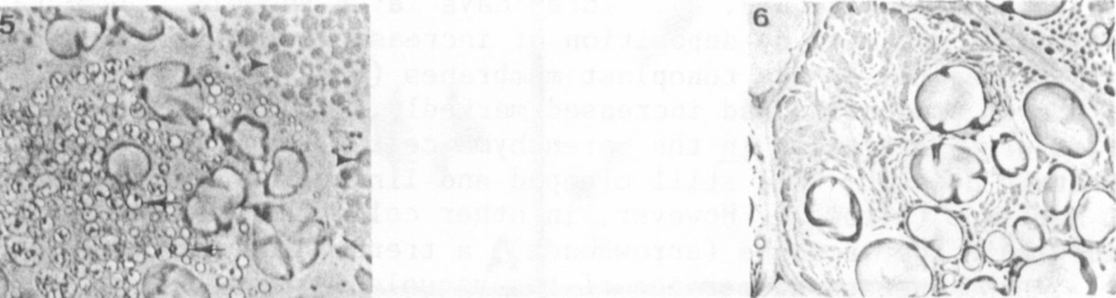
were subdivided by cytoplasmatic strands and invaginations. As shown in
the light micrograph of day 17 these invaginations led to a fragmentation
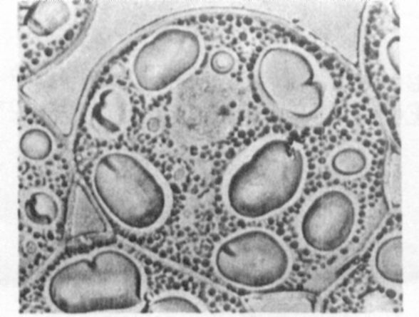
of the large vacuoles (Fig. 3). Three days later vacuole fragmentation
had begun, accompanied by deposition of increasing amounts of storage
protein aggregates at the tonoplast membranes (Fig. 4, arrows). Furth i
more, starch production had increased markedly. At day 23 numerous small
vacuoles were detectable in the parenchyma cells (Fig. 5). In some cells
the protein deposits were still clumped and lining the fragmentated tono-
plast membrane (arrows). However, in other cells the reserve protein was
spread within the vacuoles (arrowheads), a trend which continued there-
after. An homogenous dispersion of the vacuolar matrix became apparent
in the light micrograph of day 27 (Fig. 6). The morphological charac-
teristic of the cotyledon parenchyma cells at this developmental stage is
the irregular shaped appearance of the vacuolar profile. Subsequently,
spherical storage organelles, the protein bodies, were formed (Fig. 7).
The results of these light microscopic studies indicate the vacuolar ori-
gin of the protein bodies.